Imaging the mechanics of triple-negative breast cancer to advance treatment discovery
In a project bringing together an imaging specialist and biomedical expert, the team have used new imaging techniques that reveal – in unprecedented detail – what is fueling the aggressive spread of triple–negative breast cancer cells.
The challenge
Tumour stiffness – potentially key to treatment discovery – remains difficult to measure
Triple-negative breast cancers are highly aggressive, spread quickly and have limited treatment options. Each year, 2,500 women in Australia are diagnosed with this challenging disease.
Understanding tumour composition and how cancer spreads (metastasis) could enable earlier diagnosis and treatment.
However, whilst current imaging tools can see the tumour cells, they cannot “feel” the tumour properties, such as the stiffness, which is known to be important in how tumours develop, spread and respond to therapy.
Developing new imaging approaches that can see and “feel” tumour features – including variations in tissue structure, composition and behaviour – could improve early detection and treatment strategies for this challenging disease.
Using Brillouin microscopy to examine tumour tissue.
Our response
Linking tumour’s physical properties with their behaviour, using light
Overcoming this challenge required the joint expertise from imaging specialists and biomedical researchers.
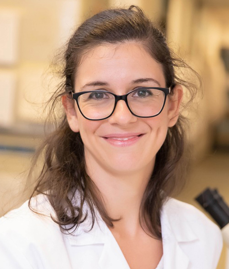
Associate Investigator Thomas Cox (Garvan Institute) and Chief Investigator Irina Kabakova (UTS) used an imaging technique – called Brillouin microscopy – to study the tumour’s mechanical properties and how that may influence the spread of tumour cells around the body.
Brillouin microscopy involves shining a laser onto different tumour regions, where it then undergoes an intricate energy exchange with the natural vibrations of the tumour tissue. This interaction creates new colours of light – like a fingerprint – revealing how stiff or soft each region is.
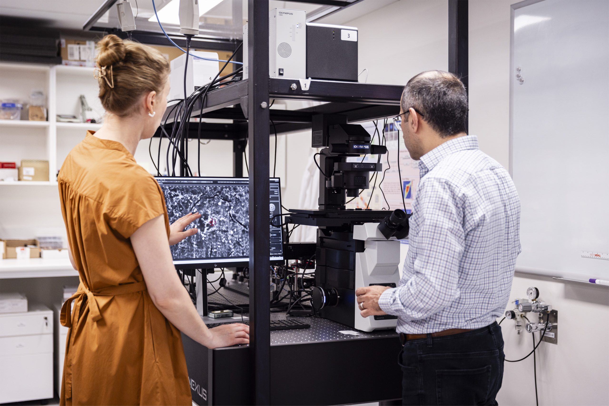
Chief Investigator Irina Kabakova and Hadi Mahmodi in the lab
The results and current progress
Starving cell’s ‘fatty diet’ of more aggressive triple-negative breast cancer cells could lead to better treatment
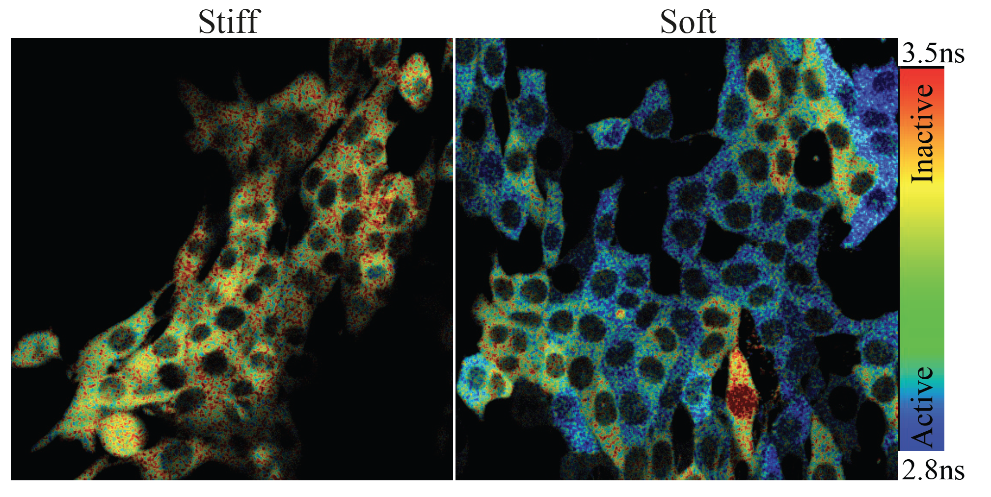
As published in Advanced Science, Brillouin microscopy revealed that triple negative breast cancer tumours are non-uniform, with both soft and stiff regions.
Crucially, the team found that a cancer cell’s location within these regions influenced its ability to spread. Cells from softer areas were more likely to spread and, unlike typical cancer cells that rely on sugars, these cells also used fats for fuel. The cell’s unique ‘fatty diet’ helped them to survive and spread.
This insight has opened new treatment possibilities, including ‘starving’ these cells of fats to limit their ability to spread.
The team now aims to explore how microcombs could speed up Brillouin microscopy – reducing processing time from hours to seconds. This could enable real-time tumour imaging to guide surgeries and preserve healthy tissue.
Read the full article in Advanced Science here.
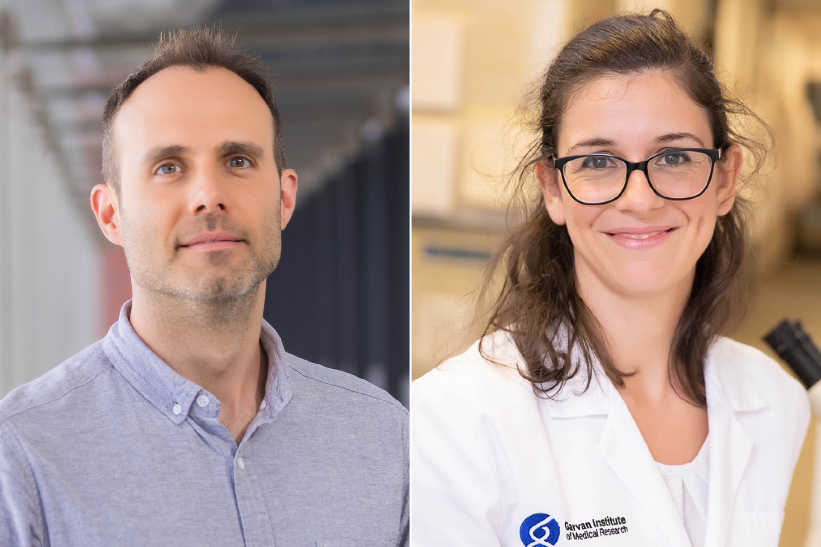
Thomas Cox and Elysse Filipe from the Garvan Institute.
