Spectroscopy and Microscopy
Grand challenge
How can optical frequency combs allow us to ‘see’ more of the world around us beyond human vision – revealing cellular structures, breath chemistry, and environmental interactions? Could they enable real-time disease detection and insight into biology – all through compact, affordable tools used anywhere in the world?
Background
The ability to ‘see’ inside the human body – first in 2D with X-rays and now in 3D with CT and MRI scans – has given experts much deeper insight into medicine and science. These techniques work because they present data as an image or 3D object that our human eyes and brains can easily interpret.
Spectroscopy takes a different approach. By measuring the specific colours of light that a sample absorbs or emits, it reveals a unique ‘fingerprint’ – providing information into the electronic state of an atom, the chemical makeup of molecules, or generally what the cell is made of.
These techniques are powerful tools across disciplines – from chemistry to biology to environmental science – but are typically limited to laboratory settings and often only capture information at a single measurement point.

What do we currently use spectroscopy for?
Spectroscopy allows us to:
- measure gases like carbon dioxide and methane in the atmosphere for tracking climate change
- identify unknown substances – such as explosives at airports, or alcohol in roadside breath tests
- assess material properties, like detecting stiff cancer cells from ‘softer’ healthy cells for earlier disease detection
What do we currently use microscopy for?
Microscopy, which forms 2D and now 3D images of samples which reveal biological structures at the microscale.
It allows us to:
- assess cell health by looking at cell’s shape and structure – for example during IVF procedures
- observe how diseases like malaria interact with blood cells, aiding treatment development
- map areas of cancerous tissue within samples to help determine the cancer stage and guide treatment decisions.

The challenge
Current imaging techniques allow us to see in very high-resolution and even in real-time, however many pieces of equipment remain slow, bulky and confined to complex laboratories, meaning they cannot be used for portable applications.
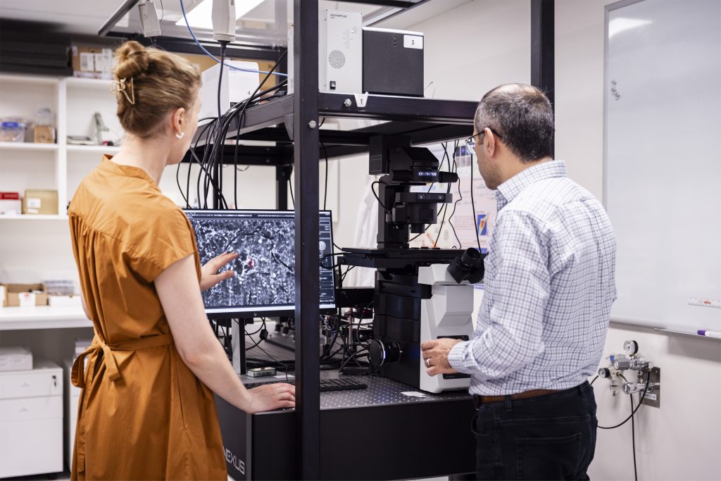
Modern microscopy – especially with the advent of light-sheet techniques – offers powerful, high-resolution 3D images of cells, which allow rich biological insight. However, while spatial imaging has advanced rapidly, combining this with chemical insight remains a major hurdle.
Spectroscopy can help – it provides rich information about a sample’s chemical and mechanical structure, but it’s slow – often requiring sequential colour-by-colour measurements – which can take many hours to get a 2D image. This limits its use in real-time or high-throughput applications.
The challenge is to dramatically speed up spectroscopic imaging using the ultra-high speed, parallel capabilities of optical microcombs. The goal is to capture an entire 2D cross-section of a sample (via a technique called light-sheet microscopy) and its complete spectrum at the same time – all in one shot. Achieving this would allow real-time, 3D visualisation of not just shape, but its underlying chemical and mechanical properties.
Ultimately, we aim to integrate this capability into next-generation microscopes that are compact, accessible and integrate multiple instruments into one – that are ultimately usable beyond high-end laboratories. This would make powerful, real-time molecular imaging much more accessible to biomedical and clinical specialists.
Our vision
Our vision is to create real-time 2D and 3D images of living systems that also capture their spectral signatures – essentially revealing chemical and mechanical changes as they happen. For example, we could ‘see’ the world in more detail than before through:
- gases being released by individual trees as they absorb carbon dioxide and emit oxygen, gaining insight into their health and life cycle.
- malaria parasites interacting with red blood cells and track cell wall stiffening happening in real-time for new treatment methods
- chemical signals passing between cells during embryo development, helping us to understand the very nature of how multi-cellular organisms form
How does our team fit into the broader Centre?
Our team works closely with:
- Microcomb Science and Technology: This theme provides photonic chips that allow us to image beyond the visible spectrum. To work effectively with biological samples and gases, we need microcombs that operate in the visible and mid-infrared – the wavelengths where water and atmospheric molecules interact more clearly than in standard telecommunications bands.
- Sensing and Measurement: We will learn from this theme’s understanding in being able to map the light spectrum of LiDAR for use in our own imaging techniques.
- Astrocombs: Our team will partner with astrocombs as we have an overlapping area of interest: sensing the atmosphere.
- Information and Intelligence: Our team will learn from the machine learning approaches to allow us to analyse large datasets of information efficiently.
Research projects

Measuring our greenhouse gases more rapidly and accurately
Our team aims to miniaturise the current bulky gas sensing equipment and use it in remote areas like Australia’s outback.
Imaging the mechanics of triple-negative breast cancer to advance treatment discovery
In a project bringing together an imaging specialist and biomedical expert, COMBS researchers have used new imaging techniques that reveal – in unprecedented detail – what is fueling the aggressive spread of triple-negative breast cancer cells.
Team

Professor Kishan Dholakia
Kishan is a biophotonics specialist with an interest in fundamental photonics and interdisciplinary science.
Associate Professor Jian Zhen Ou
Jianzhen is a specialist in developing nanomaterials and devising instruments for more accurate biological, ions, and gas sensing.

Professor Irina Kabakova
Irina is an optical physicist exploring advanced imaging techniques for modern biomedical optics, biology and bioengineering.